Cellular metabolism is a fundamental process of biology and plays an important role in cellular growth, proliferation, and differentiation. Emerging technologies that continuously monitor glucose consumption could tell us much about the health and fate of cells.
This type of information is critical for several research endeavours. For example, glucose metabolism affects the progression of diseases like cancer, says Kenan Moss, a specialist in cell and gene therapy at PHC Corporation of North America, a subsidiary of Japan-based PHC Holdings Corporation. “A key indicator of cancer is unregulated metabolic growth,” he says. While in stem cells, certain levels of metabolic activity can indicate when a cell is about to differentiate — and could soon tell us even more. “As current research progresses, we may be able to better understand how metabolic activity translates into differentiation, when that differentiation might occur, and what kind of cell lines will result,” says Moss.
A good understanding of the metabolic activity of cells in culture is important for research focused on everything from expanding our understanding of basic cellular biology, to the development of new treatments like CAR-T therapy and other cancer immunotherapies, and stem-cell-based regenerative medicines. For manufacturers of cellular products, too, good data on glucose uptake and lactate production will help optimize metabolic activity, allowing them to become more efficient, improve yields, and produce better characterized end products.
LiCellMo allows for the continuous, sample-free measurement of glucose and lactate in culture medium with results being displayed on the touch-panel controller.
Surpassing snapshots
But current methods for studying glucose metabolism leave a lot to be desired, says Moss. To get data on how metabolism changes over time, researchers must sample their cell cultures periodically, at certain times throughout the day, which introduces several problems.
First, it is labour-intensive. Laboratory staff often have to come in after hours and on weekends to collect samples at the right time. Second, it runs the risk of contaminating the cell lines, as they must be repeatedly taken out of incubation and handled to get the necessary samples. Lastly, these repeated removals can cause shock to the cell culture as they are moved from their optimal growth environments, over to sample prep areas, and back again. Researchers must balance the amount of data they need against the labour, damage, and contamination risk required to collect it.
But most importantly, no matter how often you sample the cells, you will always be missing a lot of data. Each individual sample tells you how much glucose was being used at a given time, which can be compared with the data from the next sample, but there is no information about what was going on in between those two points. “We can never take more than a snapshot,” says Moss.
Much more useful would be a way to continuously monitor the metabolic activity of cells in culture, to get a more fine-grained picture of how it changes over time. And the researchers at PHC are working on just that. “We are developing a new technology which allows real-time visualization of the metabolic condition of living cells,” says Ryosuke Takahashi, a general manager of cell and gene therapy business at PHC Corporation of North America.
The ability to monitor metabolism in real-time will immediately remove the potential for contamination and labour intensity, says Moss. The biggest benefit, however, will be the scientific discoveries continuous monitoring will make possible. It will provide both quantitative and qualitative data on not just the change in metabolic activity over time, but the rate of change of that activity, says Moss, likening it to the difference between speed and acceleration. “When you’re taking just end-point samples, you get the speed. With continuous monitoring, you get both speed and acceleration,” he says. “Instead of linear information, we can get live cellular performance rates. We can see how the glucose consumption rate changes with time.”
That will provide better insight into the activity of the cells, and their overall health, hinting at the quality of their growth by showing to what degree, and at what times, the cells are ramping up their glucose usage. For example, says Moss, periodic sampling would show that over several days the total amount of glucose taken up by a cell culture is continually increasing, because the number of cells is rising. But those periodic, discrete checks may fail to spot something that continuous monitoring would quickly make clear: that the rate of consumption over time is actually falling, indicating that individual cells are consuming less — a potential sign that the quality of their environment is deteriorating, and they are becoming less healthy. “The one-to-one ratio of how much glucose you have at the beginning versus how much you have at the end, and how big your culture is doesn’t tell you when those cells are at their most optimal,” says Moss. “But you can see that when you are looking at change over time.”
Better, more nuanced data on glycolysis will also help provide greater understanding of other closely linked but more complex cellular processes such as those in the mitochondria, as well as information on diseased cells and cancerous growth. It may also elucidate the activity of inhibitors in immunotherapy studies and what impacts changing environmental factors can have. “All of these culminate in a better understanding of cell health and performance. When we can visualize that in a more optimal way, we get a greater understanding of a lot of different processes that cascade from initial metabolism,” says Moss.
Progress at PHC
The team at PHC is taking on the daunting technical task of developing both electrochemical sensors that can work for extended periods of time in the demanding environment of a cell culture, and the software algorithms that interpret the data to provide this kind of information in a stable, reproducible way, says Takahashi. “That is the biggest challenge for us. The team is in the final stages of completing a working sensor. The hope is that this kind of system will help to open whole new worlds of metabolic research, with exciting potential for human health.”
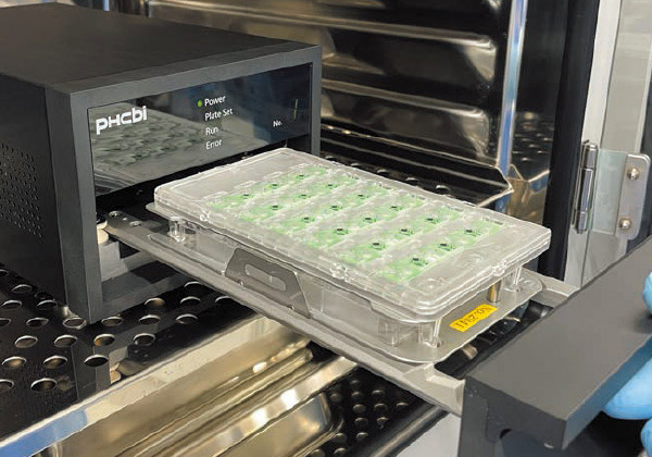
A LiCellMo sensor module attached to a common commercial plate is placed in the Detector pre-installed within the CO2 incubator.
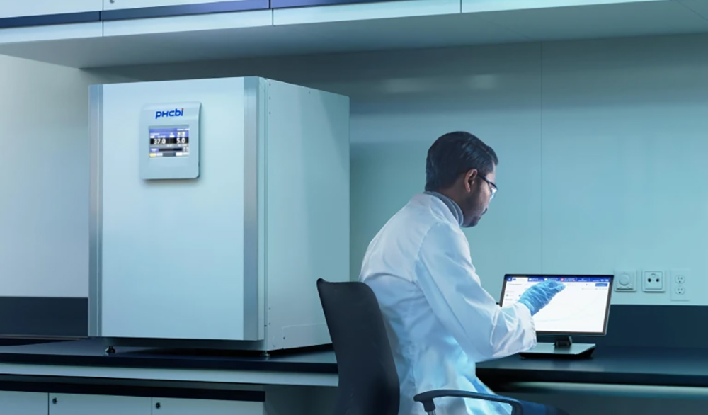
A researcher is checking the real-time measurements using the touch-panel controller.
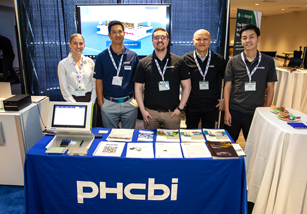
PHCbi team at a sponsored conference by Stem Cell Network in Vancouver, BC, Canada to discuss the developing new technology, LiCellMo, a live cell metabolic analyzer.
The texts contents of this article were originally produced in partnership with Nature Portfolio as an advertisement feature and published as part of Nature Index Japan in the online version of Nature (nature.com), a weekly international journal publishing the finest peer-reviewed research in science and technology.
(URL: https://www.nature.com/articles/d42473-022-00355-z)